Treating Sacroiliac Dysfunction With SI Joint Fusion
The sacrum is a triangular bone with a series of openings on either side found at the base of the spine. These holes serve as a central pass-thru for a number of essential nerve clusters. The sacrum itself serves as a vital support structure for your lower body, spine, and pelvic bone. Contrary to common perception, the pelvic bone is not a solid unit. Instead, there are joints that connect the sacrum to the iliac or hip bones. When this joint becomes compromised, it can result in a condition called sacroiliac dysfunction. A minimally invasive sacroiliac (ic) joint fusion is one method of resecuring this joint and easing chronic pain.
SI Joint Fusion Sacroiliac Dysfunction
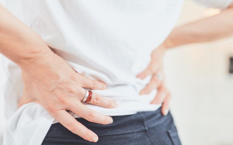
When the si joint has become unstable, it is generally referred to as sacroiliac dysfunction. Typical symptoms of this condition are pain in the lower back, legs, buttocks, groin, and feet. It is known to appear more frequently in women who have borne children due to changes to the pelvic area during pregnancy. Additional risk factors for sacroiliac dysfunction include:
- Traumatic Injury – Any traumatic injury can destabilize the pelvic area.
- Arthritis – As with any other joint, the SI joint can be impacted by the wear and tear of arthritis known as osteoarthritis. It is also subject to ankylosing spondylitis.
- Infection – Infections occurring in the SI joint can cause significant pain
- Lumbar Fusion – Studies suggest that lumbar fusion procedures may cause higher risks of sacroiliac dysfunction.
A si joint fusion may be suggested when sacroiliac dysfunction has been diagnosed and confirmed. Previously, this procedure would involve a large scar and significant recovery time. Thankfully advances in surgical technology have made a minimally invasive option available. The healing process and collateral damage from the treatment are significantly reduced.
The purpose of a sacroiliac fusion is to bond your sacrum to your iliac bones. Achieving this may involve the use of a bone graft and implanted instrumentation. The minimally invasive procedure involves the steps detailed below:
- You will be situated on your stomach on an operating table
- IV anesthetic will be provided to achieve general anesthesia
- A 1in incision will be made on one side of the buttock
- The physician will dissect the gluteal muscles to access the ilium
- A guide pin will be used in creating a small hole in the ilium
- The hole will subsequently be drilled all the way through the ilium
- This allows the implanted instrumentation tor each the sacrum
- At this point, the area will be cleared of soft tissues if a bone graft is necessary
- The bone graft will be placed at this juncture, if appropriate
- The necessary instrumentation is fitted into place
- Debris is cleared, and the area cleared prior to being closed
- The wound is closed in layers, completing the procedure
Completion of the procedure will take approximately an hour. Throughout the procedure, the physician will use fluoroscopy to guide them.
Find Out More From Your Provider
If you desire to learn more about this procedure and if it can address your concerns, speak to your spinal specialist. They’ll provide information about the procedure and assess whether it’s appropriate for your case.